


- 1
- 23
Given Footage is part of a medical school autopsy class. The body is completely dissected, the video shows the intestines being removed.

- 1
- 16
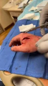
visible drainage from the pleural cavity, and if you pay attention, the doctor tried to remove the fragments by rotating them, this is because it is located opposite the inlet.


- 1
- 24
The inflammation and ulceration of the cornea. Can rapidly lead to loss of vision due to stromal degradation and thinning.

- 1
- 14
Posterior neck defect after ablative surgery. Ld flap is seen on top, before insetting. White arrows show muscles involved in neck extension and hardware.

sterilized 3d model and LdnvF prior to reconstruction. the model was useful to orientate the flap adequately.

- 1
- 21
(a) Photograph of patient with large neck abscess and hematoma following left CEA.
(b) Intra-operative photograph of ICCP.
(c) Post-operative photograph following carotid artery reconstruction, coverage with muscle flap and split skin graft.

- 1
- 22
Application of vacuum-assisted closure technique (VAC technique) in a patient with graft infection in groin
(A=Szilagyi III infection with cutaneous fistula, B=marking of infected area with use of methylene blue, C=Wound debridement and replacement of infected graft with a silver-bonded synthetic graft, D=preparation of sartorius muscle, E= coverage of the graft with the muscle flap, D=Application of VAC system
Graft preservation is reported as an alternative option, especially in high risk patients, unless there is sepsis or anastomotic bleeding. In such cases local treatment with surgical debridement, antibiotic administration and muscle flap coverage is applied. [19,30]
Treatment of peripheral grafts infection shows low mortality rates ( 0-9%) but increased amputation rates (33-67%) compared to treatment of aortic grafts infection. [11,76]


- 1
- 16


- 1
- 20


- 1
- 24
This photo shows a calcified maternal fetus that had retained in the maternal peritoneal cavity for 30 years.
Lithopedion, also known as a "stone baby," is an extremely rare medical condition that occurs when a fertilized egg implants outside the uterus and develops into a fetus. However, instead of continuing its growth within the uterus, the fetus dies and is not expelled from the mother's body. In response, the body calcifies the fetal remains to protect the mother from infection. This calcified mass, resembling a stone, is referred to as a lithopedion.
Lithopedion occurs when the fertilized egg implants in the fallopian tube, abdominal cavity, or cervix instead of the uterus. This condition typically happens due to a failed tubal pregnancy, where the fetus cannot survive or grow in the abnormal location. The mother may not be aware of the presence of a lithopedion since the body often does not exhibit any symptoms or signs of pregnancy.
Since lithopedion is a rare occurrence, estimates of its frequency are uncertain. The condition is usually discovered incidentally during medical imaging for other reasons, such as abdominal X-rays or ultrasounds performed for unrelated medical conditions.
Treatment for lithopedion involves surgical removal, as leaving the calcified mass in the body can lead to complications such as infection or pressure on surrounding organs. However, in many cases, the presence of a lithopedion is harmless and may remain undetected for a person's entire life.
It's important to note that while lithopedion is a legitimate medical condition, it is extremely rare, and most pregnancies progress normally without any complications

- 1
- 13
A 32-year-old man presented to the emergency department with a 1-month history of enlarging chest wounds.
He reported a 3-year history of daily use of fentanyl adulterated with xylazine by injection into his neck and arm veins. Physical examination showed necrotic wounds on the upper chest with exposure of the underlying clavicles (Panel A).
Computed tomography of the chest showed osteomyelitis of the clavicles and manubrium, in addition to soft-tissue ulceration and inflammation. A tissue culture grew Pseudomonas aeruginosa and enterococcus species. A diagnosis of xylazine-associated skin injury complicated by soft-tissue infection and osteomyelitis was made. Chest-wall débridement (Panel B) and microsurgical flap reconstruction (Panel C) were performed.
Intravenous antimicrobial therapy was initiated on admission and continued for 6 weeks postoperatively. Buprenorphine therapy was initiated to treat the patient's opioid-use disorder. Xylazine (also known as “tranq”) is an α2-adrenergic receptor agonist that has been approved only for veterinary use. It is an increasingly prevalent additive to fentanyl in the United States. Xylazine is associated with skin injury — commonly on the legs (Panel D, different patient) — that can occur regardless of the site of injection.
At the 6-month follow-up visit, the patient's wounds had healed well, and he had been participating in an outpatient addiction program.
- 1
- 15
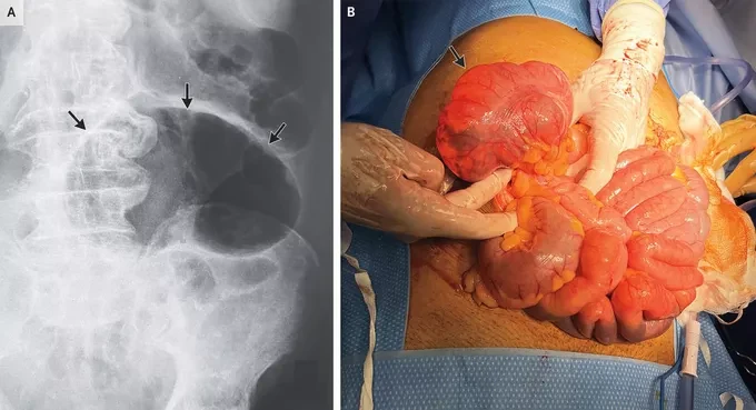
An 87-year-old man with chronic kidney disease, hypertension, and atrial fibrillation presented to the emergency department on a remote South Atlantic island where he resided with a 3-day history of constipation and lower abdominal pain. The patient's body temperature was 39.2°C, the heart rate was 97 beats per minute, the respiratory rate was 28 breaths per minute, and the blood pressure was normal.
Physical examination was notable for abdominal distention and tenderness to palpation of the right lower quadrant without rebound or guarding. A plain radiograph of the abdomen showed an air-filled loop of large bowel resembling a coffee bean (Panel A, arrows; supine view), a finding that may be seen in cecal or sigmoid volvulus. Although computed tomography of the abdomen is the definitive study in the diagnosis of bowel volvulus, it was not available at this hospital. On the basis of the available clinical data, the patient was taken urgently to the operating room. A cecal volvulus without signs of bowel compromise was identified (Panel B, arrow) and successfully detorsed.
Owing to intraoperative hemodynamic instability, a cecopexy and cecostomy were performed rather than an ileocecectomy or right colectomy. The patient's postoperative course was uncomplicated. At 6 months of follow-up, he was doing well and declined ostomy reversal at that time.

- 1
- 22


Top Poster of the Day:
 reichsfuhrer
Deaths Today: 0
Current Registered Users: 2,107,405
 BROWSE EFFORTPOSTS
SITE GUIDE
PING GROUPS
BROWSE EFFORTPOSTS
SITE GUIDE
PING GROUPS
Medical

Aspiring medical student? Or maybe just have an interest in human anatomy? Here you will find everything from autopsies to surgeries to photos from medical journals. If it happened in the operating room or the morgue, it probably belongs here.
 Slavshit

 Sandshit